

Invest in ME Research International ME Conference 2014
IIMEC9 - Mainstreaming ME Research
Click on sections below
On 30th May, 2014 I was privileged to attend the 9th Invest in ME conference in London.
The conference opened with a brief trailer about the film "Perversely Dark" - a film produced in Norway about 2 young people with ME/CFS.
This presented a moving preview of a "must see movie" with English subtitles.
The main conference was then opened by Dr Ian Gibson.
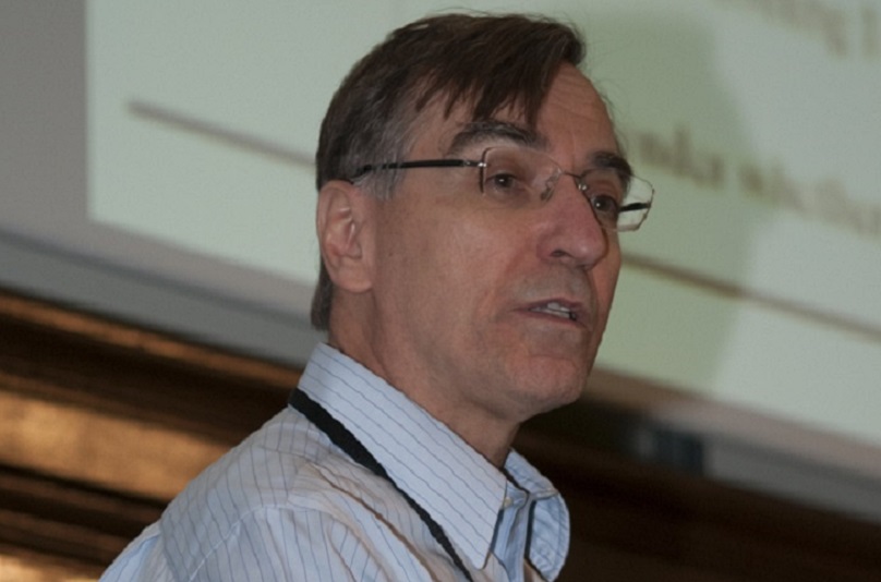
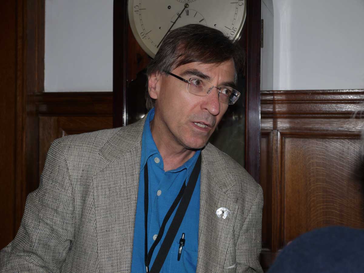
The first speaker was Prof Jonathan Edwards (London) who spoke about the lessons learnt for ME from his lifelong study of Rheumatoid Arthritis (RA). He felt he was looking from the "outside". He likened the developing studies in ME to how things had evolved with RA from 1974. The tools needed are: 1. Reproducible bio findings to build on an explanation of symptoms, and 2. A theoretical framework to build upon. By 1974, a lot had been learnt: genetic markers, an association with smoking and presence of antibodies in most patients. (Rheumatoid factors and/or anticitrulline). Inflammation is mediated by the cytokine TNF. He then asked what factors cause disease. These include: internal genetic, environmental and internal stochastic (random) - an internally driven mutation. He discussed the antigen/antibody/B cell/T cell links. The immune complex Fc gamma IIIa expresses in many tissues and leads to release of TNF. There do not have to be external triggers necessarily, but there can be self-perpetuating auto-reactive B cells.
Most auto-immunity arises by chance production of subversive B cells. The logical treatment is to remove all current B cells and start again. e.g. Rituximab - treatment with this was begun in 1998 for RA. Not all patients got better, but 2/3 had a good response - many eventually relapsed, therefore this is not the whole problem. Lessons for ME: The mechanism may be subtle. Genetic clues are like gold-dust. (e.g. NK receptors). Specific auto-antibodies make things easier, but evidence for a general immune mechanism may do. There may be no specific infective trigger, but for some with ME, maybe there is. A cytokine pathway helps. There will be several "ME diseases"- just like RA. One can be surprised by what can be achieved. In the Q&A session, he was asked if Rituximab was safe in the presence of infection. The consensus was that it should not be a problem in general, but that it should not be used in those with hepatitis C.
Angela Vincent (Oxford, UK) spoke about the searches for antibodies in neurological diseases and posed the question as to whether they could be similar to what may be happening with ME. She talked first about the classical auto-immune disease Myasthenia Gravis (MG) This is characterised by weakness and fatigue and is due to an acetylcholine defect. Acetylcholine is produced at the axon to the axon receptor, this causing muscle fibre contraction. In MG there are not enough receptors as a result of antibodies to the protein receptors. The antibodies are made by the white blood cells (specifically the B cells) and are measureable in the circulation. The tips of all antibodies are what are different between them. They bind to protein on human cells and cause auto-immune disease. Antibodies can be transferred to mice and this will cause the same disease in the mice. Patients can improve with immunotherapies such as plasma exchange, steroids and immunoglobulins. The genes may be different in early and late onset disease. Another protein important in MG is MuSK. Antibodies to VGKC (complex proteins, associated with peripheral CNS diseases) cause illnesses with many symptoms similar to ME.
Acquired neuromyotonia was described. This is associated with a lot of twitching, muscle pains etc. and is due to an auto-immune potassium channel defect. Potassium channel proteins regulate nerve depolarisation and neurotransmitter release. Another disease described was Morvan's syndrome. This is a CNS disorder with major sleep problems- there is low melatonin production. Symptoms continue even at rest. Symptoms improve with plasma exchange and immune suppression. Limbic encephalitis another auto-immune neurological disease is associated with extreme short term amnesia. Plasma sodium may be low. The antibody LGI1 is associated with memory loss and seizures and is common in limbic encephalitis. Other diseases were described including one associated with an ovarian terratoma, resulting in an encephalopathy. In another NMDAR antibodies are driven by infection eg HSV encephalitis. Antivirals can sometimes help, but patients tend to relapse. Other conditions with auto-immune probability include narcolepsy, Tourette's, autism and PANDAS. However, the relevance of antibodies in these conditions is not yet established, and some findings may be entirely incidental.
Jonas Blomberg (Uppsala, Sweden) discussed infection-induced auto-immunity in ME. His lab uses a multiplex technique, and they are able to look at hundreds of different antibodies at a time. He described how autoimmunity is avoided in the foetus by central deletion of self-specific cells in the thymus. It is normally hard for the body to distinguish friend from foe.
Autoimmunity can do damage to the CNS and peripheral nerves. ME usually starts with an eliciting event leading to its clinical hallmarks - this can be bacterial or viral. He said we need to look at the comorbidities which may often also be auto-immune. There may be post-translational modifications or non-protein antigens in both microbes and humans, plus cross-reactive conserved microbial proteins. There are signs that ME patients have impaired mitochondrial function and this may relate to post exertional malaise and exhaustion. There may be impaired energy metabolism due to block by some antibodies which affect metabolism, such as IgA anti-pyruvate dehydrogenase. Many neurological diseases have an autoimmune basis e.g. MS, GBS, narcolepsy, Tourette's, PANDAS and acute disseminated encephalomyelitis. There are also many examples of post-infectious auto-immunity. Organisms involved may be: mycoplasma, chlamydia, EBV, CMV, Toxoplasmosis, Borrelia etc. There may be cross reaction between bacteria and viruses. It is better to look for the antibodies, rather than the microbe itself. 910 antigens have been tested in their lab. It is possible to distinguish between MS and ME. He asked the question can auto-immunity explain ME? It does need further study. Looking at the co-morbidity, several organs have auto-immune aspects, but there is increasing evidence that these co-morbidities such as IBS and FM are not auto-immune.
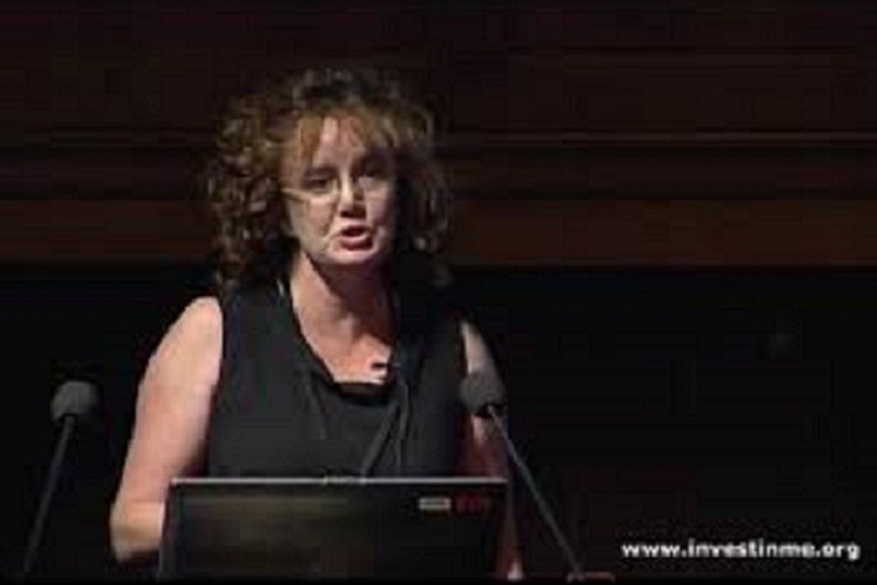
Mady Hornig (New York, USA) addressed her work on Pathogen Discovery. She has looked at the "3-Strike Hypothesis" - genes, environment and timing. Many microbes have been studied in many diseases. A lot of diseases have an immune-mediated pathogenesis. Koch postulated in 1890 that a specific microbe occurs and can be isolated in every disease. And Rivas in 1937 described the auto-immune response. Witebsky's criteria in 1957 showed that freshly circulating or cell-bound antibodies and their specific antigen target could be identified. Rose and Bona in 1993 showed that auto-antigen specific T cells may induce disease. In 1996 Fredericks and Relman demonstrated molecular markers. The blood brain barrier (BBB) is a protective lining, but it may not protect the circum-ventricular organs (CVO). There are many signs suggestive of an auto-immune response in ME, and many auto-antibodies target the brain. They may break down the BBB and get access to the CVO regions. She went on to describe as an example, PANDAS - a bacterial auto-immune neuropsychiatric disorder. There may be associated OCD, anxiety and tics. Potential pathogens implicated in ME were discussed. She described "de-discovery" of certain pathogens, such as XMRV and bornaviruses in ME as being non-implicated.
She also explained that there is now no proven link between measles vaccination and autism. However in many diseases there may be severe intestinal dysbiosis and microbiota changes may be relevant. There are a number of staged strategies of ongoing ME studies looking for pathogens. Searches for DNA and RNA agents has as yet found very little. The serum is often viral free, so there is need to look at the PBMCs. When looking for pro-inflammatory cytokines, allergy-related immune signatures are more prominent. There is decrease in ecotaxin, while many cytokines are increased. Auto-immune disturbances may result from failed uptake of dietary precursors of antioxidants in the terminal ileum. Microbiota have an important role in the tryptophan degradation pathway, and also melatonin production is affected. And auto-immune disturbances may relate to the GI tract. i.e microbes help the brain along through tryptohan and serotonin.
Carmen Scheibenbogen (Berlin, Germany) discussed the role of EBV in ME. She described how a subset have disease onset associated with EBV. Then there may be recurrent fever and nodes and the patient describes the illness as if infection is ongoing. There may be EBV IgM and EA-IgG elevation. EBV DNA is detectable in the blood. Some patients will improve with anti-viral treatment. EBV belongs to the Human Herpes Virus family, and the infection may be so mild as to be described like a common cold. The infection is lifelong and may be latent. The primary infection is usually in childhood and spread by saliva in 80% of cases. The illness is often severe in adolescence. There has been shown to be possible association with late onset EBV and auto-immune diseases such as MS and SLE. 98% adults carry latent infection. This can reactivate in immunodeficiency illnesses causing chronic active EBV, lymphoma etc. Diagnosis is by detecting specific antibodies (IgM early and IgG later).
She is currently looking at 2 projects: 1. Characterisation of EBV specific B and T cell response. She has found EBV specific antibodies: elevated EBV-IgM (marker for reactivation), absent EBV-EBNA antibodies in some with ME, diminished or absent EBV-specific memory B cells in many ME patients. These findings may indicate a deficient response due to late EBV infection or possibly frequent reactivation. Elevated EBV copies (EBER) was found in the blood of less that 10% of patients. There was no evidence for lytic replication. 2. EBV sero-array- looking at 5292 peptides. There was a different response in different patient cohorts. There was enhanced antibody response against EBV peptides in ME versus healthy controls. This is all a basis for development of diagnostic tests and treatment development.
Prof Simon Carding (Norwich, East Anglia) looked at the role for leaky gut and intestinal microbiota in the pathophysiology of ME. There has been an explosion of interest in the last 2 years. The gut is 9 metres long and has the largest collection of neural cells in the body. It could be described as our "second brain". It is also the largest immune system in the body with a huge area of surface villae. There are multi-layers of protection. The microbiota form a protective barrier. There are 100trillion microbes in the gut ranging from bacteria to fungi to viruses. So 99% of our DNA is microbial in origin. The microbiome refers to the genes. The microbiota weigh 1 kg and have a volume of 1L. There are between 300 and 1000 species. Food is the fuel for the bacteria and 1.4L of gas is produced daily. 60% of the stool is bacteria. The food and who we are shapes our microbiota. It is strongly influenced by species and region. The microbiota originate from our mothers and there are changes with age. They are there for protective function, structural function and metabolic function. In fact the intestine is a "bioreactor", and the microbiota are essential to providing our daily needs. The absence of microbiota compromises our health. "Germ free" animals have various defects as a result - such as a poor immune system and susceptibility to infection. Gut microbes however can cause disease in humans: e.g. H Pylori, clostridiae and enterococci. There is a microbiota gut/ brain axis - there is increasing evidence that the bacteria are a source of effects on brain function and disease. Stress also impacts on the microbes in the gut. The normal gut microbiota modulate brain development and behaviour. He asked the question "Is there a role involving the microbiota in ME?". There may be alterations in the intestinal barrier, leading to "leaky gut", malabsorption and inflammation. There are many possible causes of the so called leaky gut: drugs, infection, stress, antibodies, diet, neurotransmitters, cytokines, enzymes etc. Bacteria can breach a leaky barrier. There are many disease associations. In ME, IBS is common. This may be associated with auto-immune responses. Probiotics may have a potential role.
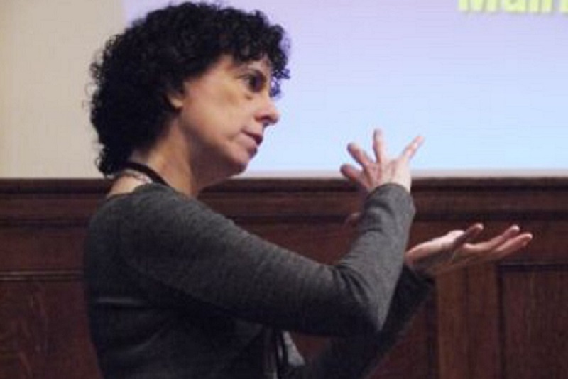
Sonya Marshall-Gradisnik (Gold Coast, Australia) updated us on the current knowledge of immunological biomarkers in ME. She initially described the different cells in the innate (dendritic and NK cells) and adaptive (NKTcells, T cells, B cells and γδT cells) immune systems. NK cell function is apoptosis by exocytosis of perforin and granzymes. There are 2 main types of NK cells: CD56dim - whose main function is lysis, and CD56bright whose main function is to produce cytokines that activate NK cells. MiRNA controls gene expression. The aim of recent studies has been to compare changes in relation to the severity of the illness. NK lysis has been shown to be decreased markedly in severe cases compared to moderate cases and controls. KIR receptors are inhibitory. The dim phenotype KIR2DL1 is significantly reduced, and CD94dim is increased in moderate and severe cases. These are responsible for increased cell lysis. Dendritic cells are increased significantly in moderate and severe cases. This is accompanied by increased production of cytokines, which cause clinical signs and symptoms. With B cell phenotypes, there is significant increase in memory and naive B cells, due to increased dendritic cell and cytokine production. This indicates an auto-immune response. γδT cell phenotypes are significantly decreased with reduced lysis function. iNKT cells are increased in severe cases and this leads to increased cytokines. NK cell lysis is low and there is significant reduction in adhesion markers. There is decreased migratory ability of NK cells to migrate towards the antigen to lyse. MiRNA plasma in ME - significant differences are expressed between ME cases and non-fatigued controls. Isolated WBCs in the plasma have an important role in the immune system in preventing inflammation, in T cell development and Treg function. In conclusion, there is consistent decrease in NK lysis and the severity of the illness reflects the immune changes. There is significant loss of function in KIRs. There is significant increase in dendritic cells, iNKTcells and naive B cell phenotypes. She has set up a cell phone APP called clinihelp and patients can access this to record symptoms regularly.
James Baraniuk (Washington, USA) discussed his work with MRI and victims of Gulf War Illness (GWI). He has explored a model sub-group dating back to 1990-91. Many had experienced acute illness, possibly as a result of sarin exposure. There has been chronic progression of illness over 20 years. Up to 25% of Gulf War veterans were affected. There are resulting problems with the brain, its functions and networks. i.e. "The pain is in the brain". This results in problems with fatigue, attention, working memory, pain, anxiety, tenderness and exertional exhaustion. Many types of MRI are now available: Voxel-based, diffusion tensor imaging, BOLD (measures oxygenation of blood in brain), fMRI (at rest and during tasks), functional connectivity, structural connectivity, cerebral blood flow and concentrations of brain chemicals. He has been interested in looking at cortical thickness, and grey and white matter intensities and lucencies. He has looked at the white matter volume and fatigue duration. The white matter is decreased in ME. There is shrinkage of 1% per year. There is also volume loss in the superior cerebellar peducles, pons and medulla in GWI. Also some brain stem atrophy. Using BOLD and fMRI, these measures are blood oxygenation level dependant and the areas that are functioning can be identified. Brain blood flow is regulated from the neuron, which releases glutamate, affecting NMDA, which then activates astrocytes to produce D-serine, which releases nitric oxide, which relaxes the arterioles and thus increases the blood flow. The fMRI shows differences in GWI leading to more errors. He then discussed resting state brain networks. When the mind is wandering, it is rehearsing and/or debriefing. A default network starts. When you stop a task, there is functional connectivity. Patterns of resting state networks may be indicative of specific diseases. He also talked about the differences in easy and difficult tasks, and also what happens when you switch tasks. Different parts of the brain function depending on difficulty. You may go into default mode if you need to switch tasks. If a person has post-exertional malaise, there are differences in cognitive function identified also. In a 2 day exercise test, healthy controls experienced some fatigue and switched to default mode, but those with GWI found symptoms came on, and there was basal ganglia activation for cognitive compensation. In a second test everything went down and additional brain areas were recruited. The GWI patients could be divided into phenotypes according to their response to exercise. There were START and STOPP groups. In the former exercise caused autonomic dysfunction. In the STOPP group there was phantom perception. He finished by saying that these new brain scanning studies will redefine psychiatry. For example, in depression the amygdala has a central role and has dysfunction, while the cerebellum is shown to be involved in emotional processing and fibres have connection with amygdala and areas of atrophy.
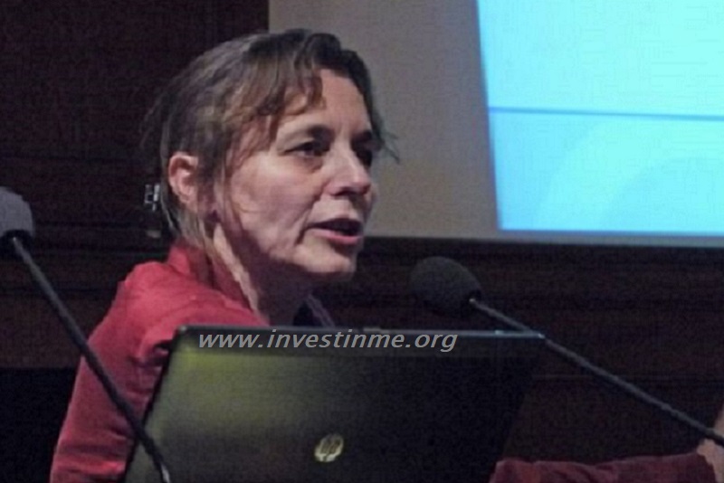
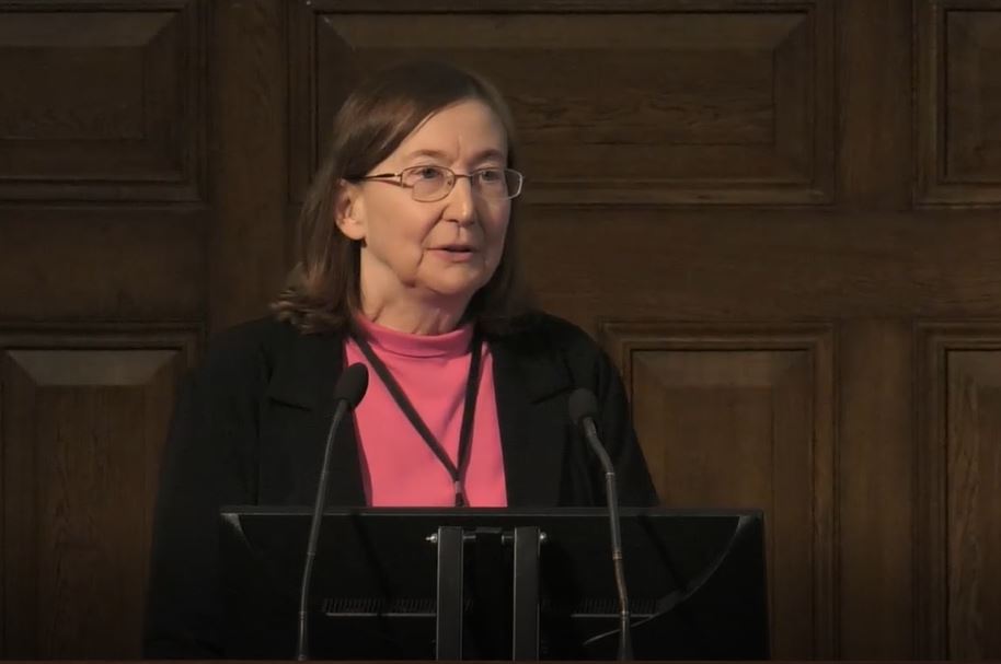
Julia Newton (Newcastle, UK) focused on the Autonomic Nervous System (ANS)and its relationship to ME. She explained that there is overlap between the ANS and many diseases associated with fatigue. The experience of fatigue is the same in many diseases. She described the ANS, and said that dysautonomia in ME is likely. The fatigue in 89% of those with ME may be due to Orthostatic Intolerance (OI). There are objective measures which can be done such as BP, HRV, tilt-table testing (HUT). Neurally mediated hypotension and POTS can thus be diagnosed. Mechanisms are upstream to the brain and downstream to the vascular system. The valsalva manoeuvre is used in some studies, particularly those associated with cognitive performance. Measurements can be taken using MR spectroscopy associated with 2 minutes of exercise. Acid accumulates in the muscles in ME. The intracellular pH has been measured in cultured muscle cells from ME patients and controls, and there was significant difference with increase in acid in those with ME after exercise. The liver is very involved in BP control, and liver volume can be measured while performing 15 seconds of valsalva. The liver volume changes dramatically. In ME there may be problems with liver volume. Using cardiac MRI, 1/3 ME patients had a significant PCr/ATP value of less than 1.6. There was exaggerated torsion of the left ventricle during pumping. These measures confirmed that there were autonomic abnormalities in ME, with associated brain, cardiac and muscle abnormalities. However there were similar findings in other fatigue related diseases. Fatigue is common and can relate to very specific physiological abnormalities. Symptoms are suggestive of ANS dysfunction. The dysfunction also correlates with fatigue severity.
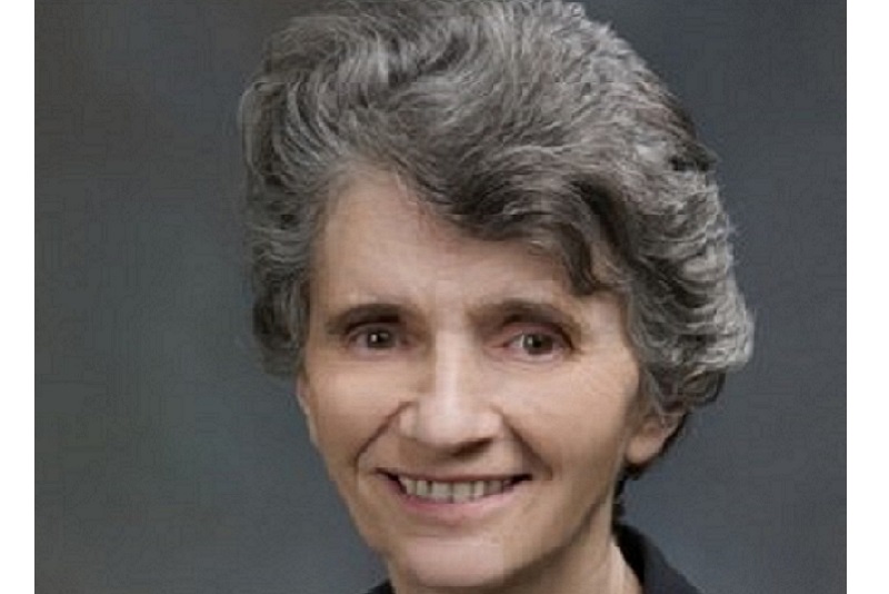
Maureen Hanson (New York,USA) - discussed markers of post-exertional malaise. She pointed out that exercise does not usually exacerbate symptoms in healthy people or in most other diseases. In ME exercise causes worsening of symptoms. CPET using a bike with resistance showed on a 2nd test 24 hours later that CPET values could not be reproduced in ME patients. In other diseases patients can usually reproduce their base response 24 hours later (e.g. heart failure, end stage renal disease). There is therefore something odd going on in ME. Other studies have also shown that having the 2nd test is important. There is a need to prove that it is not just a matter of the ME patients not trying on the 2nd test. Resting exercise rate= CO2 exhaled/O2 consumed. This will equate to equal to or greater than 1:1 with maximum effort. At rest 0.8 is typical. As effort increases, muscles release CO2 and more oxygen is consumed. VO2 max equates to the level of anaerobic physical fitness. VO2 is the volume of oxygen consumed per minute. At ventilatory threshold, (VT) anaerobic metabolism begins. Patients cannot wilfully alter the amount of oxygen they inhale or the amount of CO2 they exhale. ME patients showed a 25% decrease on VO2 max on second day. In patients who also have dysautonomia, the BP does not go up and they have to stop. Sub groups have been detected also in the 2nd CPET, which may correlate with signalling molecules in the blood. There are changes in chemokines and cytokines. 10 cytokines were measured and 5 were decreased markedly. A pilot study compared metabolites in ME patients and found 52 significant differences between before CPET1 and after CPET2. There was reduction in several acylcarnitines after exercise. 300 polar metabolites were examined and 83 differed significantly. Most were higher in controls than patients. Acetyl-carnosine was 2-fold lower in patients than controls. In conclusion: ME patients cannot reproduce their performance on a 2nd CPET. The abnormal responses can affect the autonomic or physiological responses to exercise. Both cytokines and plasma metabolites are altered compared to controls.
Amolak Bansal (Surrey, UK) discussed diagnosis and treatment of ME within the NHS. His initial comments mentioned that eventually the current exclusion criteria may go on to be included, and that if anything the new ICC can make things more complicated. His team use the Sutton CFS/ME scoring system, needing 8 out of 13 points to make a diagnosis. He noted in particular that if a patient can manage 4 glasses of alcohol in one sitting he is unlikely to have ME (usually there is extreme sensitivity to alcohol). When comparing ME to depression, there is more motivation coupled with often adverse reactions to anti-depressants. Conditions which can mimic ME include: joint hypermobility, hypothyroidism, Addison's disease, gluten sensitivity, Sjogren's syndrome, primary sleep disorders, cardiac disease, Parkinson's disease, persistent anxiety and depression. When examining an ME patient there is an abnormality in the pupils. Holding the light there, there will be constriction - dilation and then further constriction. Other signs include increased respiratory rate and cold peripheries. Vitamin D should be checked as there is risk of osteoporosis. There is little or no evidence of fungal infection. Treatment plan should include: stress management, gentle exercise and sensible diet. B12 injections can help with cognitive symptoms. Other supplements of use include: magnesium, L-carnitine, CoQ10 and D-Ribose. Naltrexone and nimodipine help some patients. Hormones such as thyroid, growth hormone, glucocorticoids and oestrogen may be appropriate for some people. Other treatment options to consider include: immunotherapy, antivirals, antibiotics, ampligen and anti-B cell therapy. Betablockers can be useful for anxiety.
Andreas Kogelnik (California,USA) went on to discuss the diagnosis and treatment of ME in the USA. He stressed that this is not a psychiatric disease. He outlined the many activities of the Open Medicine Institute. They are gathering very big data. As ME is a multi-system disease, many different methodologies are needed. For some patients there are three and a half billion data points. But a huge amount of time would be needed to analyse it all. He then outlined some of their current studies. These include: 1. Proteomics - 64 patients in 4 subgroups looking at autoantibody arrays. Already EBV is featuring prominently. 2. Genetics - MTHFR - so far mutations are disproportionately represented in ME. 3. Large multisite ME study 4. Gene expression profiling 5. Functional gene classes 6. Viral studies 7. Exercise testing 8. Treatment pilots which include: antivirals, IV immunoglobulin, rituximab, metabolic pathways.
Julian Blanco (Barcelona,Spain) gave an external view of ME research strategies. He compared the number of papers written for HIV with those for ME. There were many more for HIV. Research priorities tend to look at other fields - such as cancer, AIDS, neurodegenerative diseases and cardiovascular diseases. ME is a social problem with lower visibility, an economic problem (but there is more data on other diseases) and a scientific challenge with no clear target. The situation needs to be reversed. It needs more money, social visibility, and pressure on policy makers. The latter should include epidemiological data and economic impact. Biomedical research can offer: genomics, proteomics, imaging cell function (flow cytometry), B cell function and systems biology. These can all help to unravel the complexities. There is need for well defined, large study populations. Hard clinical work is also needed. The required logistics include sample storage, data management and multi-disciplinary approaches. Regarding treatment, the example of rituximab should be followed. There should be no treatment without clinical basis, and treatment should be done in a clinical trial setting. It is the patients who are moving the treatment ahead. His concluding words were: "Dialogue between science and society has never been more important". The conference was closed by Dr Ian Gibson who reiterated these comments. I must thank ANZMES and Invest in ME for making it possible for me to attend this worthwhile event. Things are moving forward rapidly, and while much work lies ahead, the new directions and science have become increasingly exciting.
Conference Presentations from IIMEC9


Images from IIMEC9, London, 2014


Bansal Score Chart
Journal of IiME Volume 8 Issue 1
IIMEC9 Pre-Conference Dinner Speech

IIMEC9 Pre-Conference Dinner Speech from Dr Nigel Speight
BRMEC4 - International Research Colloquium, London 2014

Invest in ME organised and hosted the Biomedical Research into ME Colloquium
number 4 in May 2014.
Almost fifty researchers from eight countries attended a full day meeting.
This is now an important fixture in the research calendar and a
unique event for ME.
Use this link to go to the BRMEC Colloquium page.
Our Sponsors for IIMEC9
Again the Irish ME Trust continued to sponsor a speaker for our international conference and we would like to thank them for their wonderful support.
Dr Nigel Speight
Dr Nigel Speight
Dr Nigel Speight was a consultant paediatrician in Durham for over 25 years. He has seen a large number of cases of childhood ME in his own area and has frequently been called on to support cases of where children have been treated poorly by social and healthcare services. He has played a major role in rescuing children from care proceedings and is well qualified to comment on the state of treatment of ME patients. Dr Speight presented at the 2nd Invest in ME Research International ME Conference 2007 in London and gave the pre-conference dinner keynote speech at the 9th Invest in ME Research International ME COnference in 2014. He is considered to be one of the most experienced ME consultants in the UK.
Further Information
Read more of the The General Medical Council - Dr Nigel Speight
- click here
Dr Ian Gibson
Coming Soon
Former Dean of Biological Sciences, UEA
Dr Ian Gibson was the former Labour MP for Norwich North. Dr Gibson worked at University of East Anglia for 32 years,
became Dean of the School of Biological Sciences at UEA in 1991
and was head of a cancer research team and set up the Francesca Gunn Leukaemia Laboratory at UEA.
In 2011 Dr Gibson received an honorary doctorate of civil law from UEA.
A scientist, politician and academic - Dr.Ian Gibson was uniquely qualified to comment on how science and politics have become intertwined.
Other Links
-
References
Professor Jonathan Edwards
Professor Jonathan Edwards
Emeritus Professor of Connective Tissue Medicine, UCL, UK
Professor Angela Vincent
Professor Angela Vincent
Emeritus Professor of Neuroimmunology, University of Oxford
Professor Vincent is Emeritus Professor of Neuroimmunology at the University of Oxford, and an Emeritus Fellow of Somerville College. She holds an Honorary Consultant position in Immunology and runs the Clinical Neuroimmunology service which is an international referral centre for the measurement of antibodies in neurological diseases.
Together with colleagues she collaborates with neurologists worldwide. She was formerly Head of Department of Clinical Neurology (2005-2008), and is a Past President of the International Society of Neuroimmunology, and an Associate Editor of Brain.
She was a co-applicant and group leader of OXION, the Wellcome Trust-funded Integrative Physiology Initiative "Ion channels and Diseases of Electrically Excitable Cells". She is a member of Faculty of 1000 (Neuroscience, Neurobiology of Disease and Regeneration)
Her major interest is in the role of autoimmunity in neurological diseases, including multiple sclerosis and auto-antibody mediated ion channel and receptor disorders. Recent advances have included (a) the discovery that maternal antibodies to different fetal proteins can cause rare neuromuscular disorders, and may be involved in some forms of autism or other neurodevelopmental disorders; (b) the definition and characterisation of a new form of myasthenia gravis associated with antibodies to a receptor tyrosine kinase, MuSK, that performs an important maintenance role at the neuromuscular junction; and (c) the recognition that some central nervous system disorders, involving memory loss, seizures, movement disorders, can be caused by antibodies to potassium ion channels and to various receptor proteins.
In these, and several other conditions, new ways are being devised to measure the pathogenic antibodies for better clinical diagnosis, and establishing model in vitro and in vivo systems for investigation of the pathophysiology of the diseases. Her group also works, in collaboration with Profs David Beeson and Nick Willcox, on the genetics of myasthenia and the factors that determine autoimmune responses to the main target, the acetylcholine receptor.
Other Links
Professor Jonas Blomberg
Professor Jonas Blomberg
Emeritus Professor of Clinical Virology, Department of Medical Sciences, Uppsala University, Sweden
Professor Jonas Blomberg is an MD and PhD, graduating at the University of Gothenburg. Has worked with Lipids at the department of Medical Biochemistry 1965-1972 as a Clinical Virologist in Gothenburg 1972-1979 and as a postDoc at John Stephensons Lab at NCI Frederick on retroviruses 1979-1981. He then worked as a Clinical Virologist in Lund, Sweden 1981-1995 and then as a professor of Clinical Virology in Uppsala 1996- to the present.
His main fields of interest are: Retrovirology, Bioinformatics, Clinical Virology and broadly targeted and multiplex methods for detection of microbial nucleic acid.
He also is interested in evolution and Infection biology.
Professor Blomberg is on the editorial board of Journal of Virology http://jvi.asm.org/site/misc/edboard.xhtml.
Other Links
Assoc.Professor Mady Hornig
Mady Hornig, MA, MD did her undergraduate studies as a College Scholar at Cornell, received an MA in Psychology from The New School for Social Research and an MD from The Medical College of Pennsylvania and completed her residency in psychiatry at The Medical Center Hospital of Vermont and an NIMH/NRSA Neuropsychopharmacology Fellowship at the University of Pennsylvania. Her research leverages large epidemiologic cohorts, novel bench science and animal model studies to determine how microbial, immune and toxic exposures impact upon the brain across the life course, resulting in disorders such as autism, attention-deficit/hyperactivity disorder (ADHD), Pediatric Autoimmune Neuropsychiatric Disorders Associated with Streptococcal infection (PANDAS), mood disorders, schizophrenia, myalgic encephalomyelitis/chronic fatigue syndrome (ME/CFS) and age-related cognitive deficits.
Dr. Hornig is internationally known for her work in the growing research arena exploring the mechanisms of gut-immune-brain axis functioning, seeking clues to both the understanding of the roots of dysfunction as well as uncovering pathways that strengthen individual resiliency. She has a keen interest in how diet, exercise and environmental factors affect each individual’s intestinal bacteria – the so-called gut microbiome – which then influences brain function through alterations in blood-borne molecules.
She has identified naturally-occurring substances that appear to strengthen resistance to certain disease states affecting the brain, and is pursuing these as candidates for prevention and intervention in ME/CFS and autism. She uses immune profiling, metabolomic, proteomic, epigenetic and microbiome approaches to identify prenatal and birth biomarkers for brain disorders in large prospective studies in Scandinavia as well as the US. She is also applying these approaches to uncover markers of disturbed immunity and metabolism correlating with the severe clinical deficits that underlie ME/CFS, work launched with support from the Hutchins Family Foundation/Chronic Fatigue Initiative, the National Institutes of Health and the crowd-funding initiative, The Microbe Discovery Project. Perhaps most exciting is that new ME/CFS subsets that appear to have different triggers and may respond differentially to treatment are now being identified through her work.
Dr. Hornig’s approach is enriched by her unusual combination of decades of experience as a clinical researcher, her acumen in defining novel neuropharmacological and nutritional approaches for brain disorders and her ability to carefully tease out factors that enhance resiliency to disease.
In 2004, Dr. Hornig presented to the Institute of Medicine Immunization Safety Review Committee and testified twice before congressional subcommittees regarding the role of infections and toxins in autism pathogenesis and has lectured on ME/CFS throughout the world. She has over 120 peer-reviewed publications, has edited several books, and has received many academic awards. Her work has been featured by the New York Times, the Los Angeles Times, The Washington Post, The Wall Street Journal, The Atlantic, Discover Magazine, Nature Medicine, Science, Wired, the Huffington Post, O Magazine, CBS News, and This Week in Virology.
-
References
Professor Simon Carding
Upon completing postgraduate work at the Medical Research Council’s Clinical Research Centre in Harrow, Professor Carding “emigrated” to the USA to take up a postdoctoral position at New York University School of Medicine, and then at Yale University as a Howard Hughes Fellow in the Immunobiology Group at Yale University with Profs Kim Bottomly and Charlie Janeway Jr. While at Yale an interest in gamma-delta (γδ) T cells was acquired working closely with Adrian Hayday on molecular genetics and then with Prof. Peter Doherty to establish their role in (viral) infectious disease.
He left Yale after five years to take up a faculty position at the University of Pennsylvania in Philadelphia where he developed a research interest in mucosal and GI-tract immunology, performing studies in germfree mice with Prof John Cebra that helped establish the role of gut microbes in the aetiology of inflammatory bowel disease (IBD).
After 15 years in the USA, he returned to the UK to take up the Chair in Molecular Immunology at the University of Leeds where he established a new research programme on commensal gut bacteria and Bacteroides genetics leading to the development of a Bacteroides drug delivery platform that is being used for developing new interventions for IBD and for mucosal vaccination.
In 2008 he was recruited by UEA and IFR to develop a gut research programme, taking up the Chair of Mucosal Immunology at UEA-MED and the position of head of the Gut Biology Research Programme at IFR, which later became part of the Gut Health and Food Safety (GHFS) Programme.
GHFS research covers a broad area of gut biology including epithelial cell physiology, mucus and glycobiology, mucosal immunology, commensal microbiology, foodborne bacterial pathogens, and mathematical modelling and bioinformatics. The success of this programme has led to the establishment of the Gut Microbes and Health research programme that is integral to the research agenda of The Quadram Institute.
-
References
Professor Carmen Scheibenbogen
Professor Carmen Scheibenbogen
Full professor and Deputy Chair at the Institute of Medical Immunology, Charité
Head of the Outpatient Clinic for Adult Immunodeficiencies at Charité Hospital, Berlin, Germany.
Head of Immunodeficiency Outpatient Clinic | Specialist in hematology, oncology and specialist immunologist
Professor James Baraniuk
Professor of Medicine at Georgetown University Medical Centre, washington, USA
James N. Baraniuk was born in Alberta, Canada, south of Banff. He earned his honours degree in chemistry and microbiology, medical degree, and
unique bachelor's degree in medicine (cardiology) at the University of Manitoba, Winnipeg, Canada.
Thereafter, he moved to Akron, OH, USA, for his internship and internal medicine residency at St Thomas Hospital.
After another year of internal medicine residency at Duke University Medical Center, Durham, NC, he trained with Dr C.E. Buckley, III, in allergy and clinical immunology. He moved to the laboratory of Dr Michael Kaliner at the National Institute of Allergy and Infectious Diseases, Bethesda, MD, and there began his long-standing collaboration with Dr Kimihiro Ohkubo.
After 2 years studying neuropeptides, he joined Dr Peter Barnes' laboratory at the National Heart and Lung Institute, Brompton Hospital, London, UK. Dr Baraniuk returned to Washington, DC, and Georgetown University, where he is currently Associate Professor with Tenure in the Department of Medicine.
-
References
Professor Julia Newton
Dr Julia Newton - Institute of Cellular Medicine, Newcastle University, UK
Dr Julia Newton is Senior Lecturer at the Institute of Cellular Medicine, Newcastle University. Dr Newton has been working on autonomic dysfunction in ME/CFS patients and will be presenting results of her continuing research.
Professor Maureen Hanson
Department of Molecular Biology and Genetics, Cornell
Maureen Hanson is Liberty Hyde Bailey Professor in the Department of Molecular Biology and Genetics at Cornell University in Ithaca, NY.
Previously she was on the faculty of the Department of Biology at the University of Virginia in Charlottesville and an NIH NRSA postdoctoral fellow at
Harvard, where she also completed her Ph.D. degree.
While most of her prior research has concerned cell and molecular biology in plant cells, she began a research program on ME/CFS after noting at a
2007 IACFS meeting the paucity of molecular biologists studying the illness.
Her lab was part of the 2012 multicenter study organized by Ian Lipkin's group at Columbia University to assess the actual role of XMRV in ME/CFS.
Professor Sonya Marshall-Gradisnik
Professor Sonya Marshall-Gradisnik NCNED, Australia
Co-director of the National Centre for Neuroimmunology and Emerging Diseases (NCNED) at Australia’s Griffith University. Professor Sonya Marshall-Gradisnik is one of Australia's foremost researchers in the area of neuroimmunology and has been instrumental in establishing the Public Health and Neuroimmunology Unit (PHANU) at Bond University. Much of her work relates specifically to autoimmunity in Chronic Fatigue Syndrome sufferers and she is regularly asked to speak to community groups on behalf of Queensland Health and NSW Health. Her research in the area of exercise immunology has also contributed to the body of knowledge relating to the effect of doping in sport and she serves as Sports Medicine Australia's national spokesperson in this area. The vital research conducted by Professor Marshall has attracted more than $1 million in grant funding and she has produced 21 peer-reviewed papers, five book chapters and one provisional patent. In 2008 Dr Marshall was joint leader of the Bond University team responsible for developing the the BioSMART program. The team was awarded a prestigious Australian Teaching and Learning Council Award (formerly known as the Carrick Award) for Outstanding Contribution to Student Learning and for the quality of student learning over a sustained period of time.
Dr Andreas Kogelnik
Dr Andreas Kogelnik
Dr Andreas Kogelnik is the Founding Director of the Open Medicine Institute, a collaborative, community-based translational research institute dedicated to personalized medicine with a human touch while using the latest advances in medicine, informatics, genomics, and biotechnology.
The Institute works closely with the Open Medicine Clinic and other clinics to conduct research and apply new knowledge back into clinical practice.
Dr. Kogelnik received his M.D. from Emory University School of Medicine in Atlanta and his Ph.D. in bioengineering/bioinformatics from the Georgia Institute of Technology. Subsequently, he completed is residency in Internal Medicine and a Fellowship in Infectious Diseases at Stanford University and its affiliated hospitals. Following his clinical training, he remained at Stanford with NIH funding to engage in post-doctoral research in microbiology, immunology and bioinformatics with Dr. Ellen Jo Baron and Dr. Stanley Falkow, where he explored host-response profiles in severely ill patients.
Together with Dr. José Montoya, he was instrumental in the conception, design, and execution of the EVOLVE study - a placebo-controlled, double-blind study of a subset of chronic fatigue syndrome patients with evidence of viral infection.
Dr. Kogelnik worked with Dr. Atul Butte in translational informatics to determine patterns that indicated a high risk for adverse events in paediatric patients at Lucille Packard Children's Hospital.
H
e is the Medical Director of the Open Medicine Clinic - a community-based research clinic focussed on chronic infectious diseases, neuroimmune disease, and immunology. Dr. Kogelnik has published numerous scientific papers and book chapters, is an Editor of Computers in Medicine and Biology, and is a Consulting Assistant Professor at Stanford University. With the Open Medicine Institute, he has led the formation of CFS and Lyme Registries and Biobanks as well as creating an infrastructure for providers to collect better data and implement clinical trials across a network of sites.
Dr Amolak Bansal
Dr Amolak Bansal
Consultant Immunologist St Helier University Hospitals, NHS Trust
Dr Julian Blanco
pending
Professor Jonathan Edwards
Emeritus Professor of Connective Tissue Medicine, UCL, UK
IIMEC9 Plenary Session
Coming Soon
Error processing SSI file