Summary
Background:Q fever is a common and
acute but rare chronic zoonosis caused by Coxiella
burnetii. Its acute form manifests as atypical
pneumonia, flu-like syndrome, or hepatitis. Some authors
observed symptoms of chronic fatigue in a small number
of patients after the acute phase of Q fever; in many
cases serological assay confirmed the activity of
Coxiella burnetii infection. The effect of
antibiotic therapy on post-Q-fever fatigue syndrome has
not been studied in south-east Europe thus far.
Case Reports: Three patients are
presented with post-Q-fever fatigue syndrome. All
fulfilled the CDC criteria for chronic fatigue syndrome.
IgA antibodies to phase I of the growth cycle of
Coxiella burnetii were positive in two patients and
negative in one. Two patients were treated with
doxycycline for two weeks in the acute phase of illness
and one with a combination of erythromycin and
gentamycin.
After 4–12 months they developed
post-Q-fever fatigue syndrome and were treated with
intracellular active antibiotics (fl uoroquinolones and
tetracycline) for 3–12 months. Effi cacy of the
treatment was observed in two patients, but in one
patient the results were not encouraging.
Conclusions: These results suggest the possibility of
the involvement of Coxiella burnetii infection in
the evolution of chronic fatigue syndrome. This is the
fi rst report on post-Q-fever fatigue syndrome in
Mediterranean countries. Evidence of IgA antibodies to
phase I of the growth cycle of Coxiella burnetii
is not a prerequisite for establishing a diagnosis of
CFS. The recommendation of antibiotic treatment in
post-Q-fever fatigue syndrome requires further
investigation.
| keywords: |
chronic fatigue
syndrome • Coxiella burnetii • post-Q fever fatigue
syndrome • antibiotic treatment |

BACKGROUND
Q fever is one of the most common
anthropozoonoses in southeast Europe. It is caused by Coxiella
burnetii, an intracellular pathogen whose classifi cation has
been changed from the order of Rickettsiaceae to the order of
Legionellales [1]. Human infection develops after inhalation
of contaminated aerosol or consumption of unpasteurized milk. It is
rarely transmitted by vectors, transfusions of contaminated blood,
or transplancentally [2,3]. Recently, a major role in disease spread
was attributed to air currents [4]. About 60% of infections caused
by Coxiella burnetii are asymptomatic [2]. Acute infection
usually presents as a febrile state, pneumonia, or hepatitis, while
other organs are less commonly affected. Coxiella burnetii is
endemic in rural, coastal, and non-coastal areas of southern Croatia
and is associated with stockbreeding. Acute Q fever in
Split-Dalmatia County (470,000 inhabitants) is most commonly
presented with both pneumonia and hepatitis (60.0%), followed by
pneumonia (25.8%), hepatitis (9%), and nonspecific febrile illnesses
(5.2%). During the period from 1985 to 2002, 155 acute Q fever cases
were hospitalized at the Split University Hospital, with a mean
annual incidence of 1.82/100,000/year. All cases were verifi ed by
serologic testing with C. burnetii phase II antigen as is
routinely done in all patients with clinical syndrome of atypical
pneumonia that live in endemic areas [5]. In the northern part of
Croatia, Coxiella burnetii causes 6.45% of all interstitial
pneumonias that are serologically verified [6].
In its chronic form, Q fever mostly presents
as endocarditis, infl ammation of intravascular implants,
osteoarthritis, and chronic hepatitis [7]. During a follow-up of
convalescent patients after acute Q
we noticed that some had
symptoms that were consistent with chronic fatigue syndrome (CFS).
The diagnostic criteria for CFS include fatigue for six months or
more together with at least four of the following symptoms: lack of
concentration or/and memory that interferes with normal activities,
sore throat, tender cervical or axillary lymph nodes, joint pain
without swelling, muscle pain, headache, no refreshing sleep, and
malaise lasting longer than 24 hours after exertion [8]. CFS is
twice as common in females as in males, and it is most common
between 25–45 years of age. The cause of CFS is not fully
understood. There are three hypotheses about the cause of this
impairment: postinfectious, immunological, and depression [9,10].
Penttila and associates found that in Australia, 20% of patients
after acute Q fever develop post-Q-fever fatigue syndrome (QFS).
Increased concentrations of IL-6 and interferon- as well as lowered
concentrations of IL-2 that are found after stimulating peripheral
blood mononuclear cells in cultures from these patients are presumed
to be implicated in the pathogenesis of QFS [11].
The purpose of this paper is to
emphasize the existence of CFS after Q fever in Croatia and its
incidence and to show the effects of antimicrobial therapy of
patients with QFS. We describe three patients who had QFS. During
the period from January 2000 to December 2004, 90 patients with
acute Q fever were treated at the Split University Hospital and we
observed 3/90 patients with post-Q-fever fatigue syndrome. After the
diagnosis of QFS was established, these patients were treated with
antibiotics. They were asked to fill out questionnaires assessing
their clinical condition before and after the treatment. The
questionnaire survey included subjective symptoms: fatigue, lack of
concentration, no refreshing sleep, sore throat, tender cervical or
axillary lymph nodes, joint pain without swelling, muscle pain,
headache, and malaise lasting longer than 24 hours after exertion.
These symptoms were evaluated according to four grades (0: absent,
1: mild, 2: moderate, 3: severe). If the summed result of the survey
was halved after the treatment, the effect of antibiotic therapy was
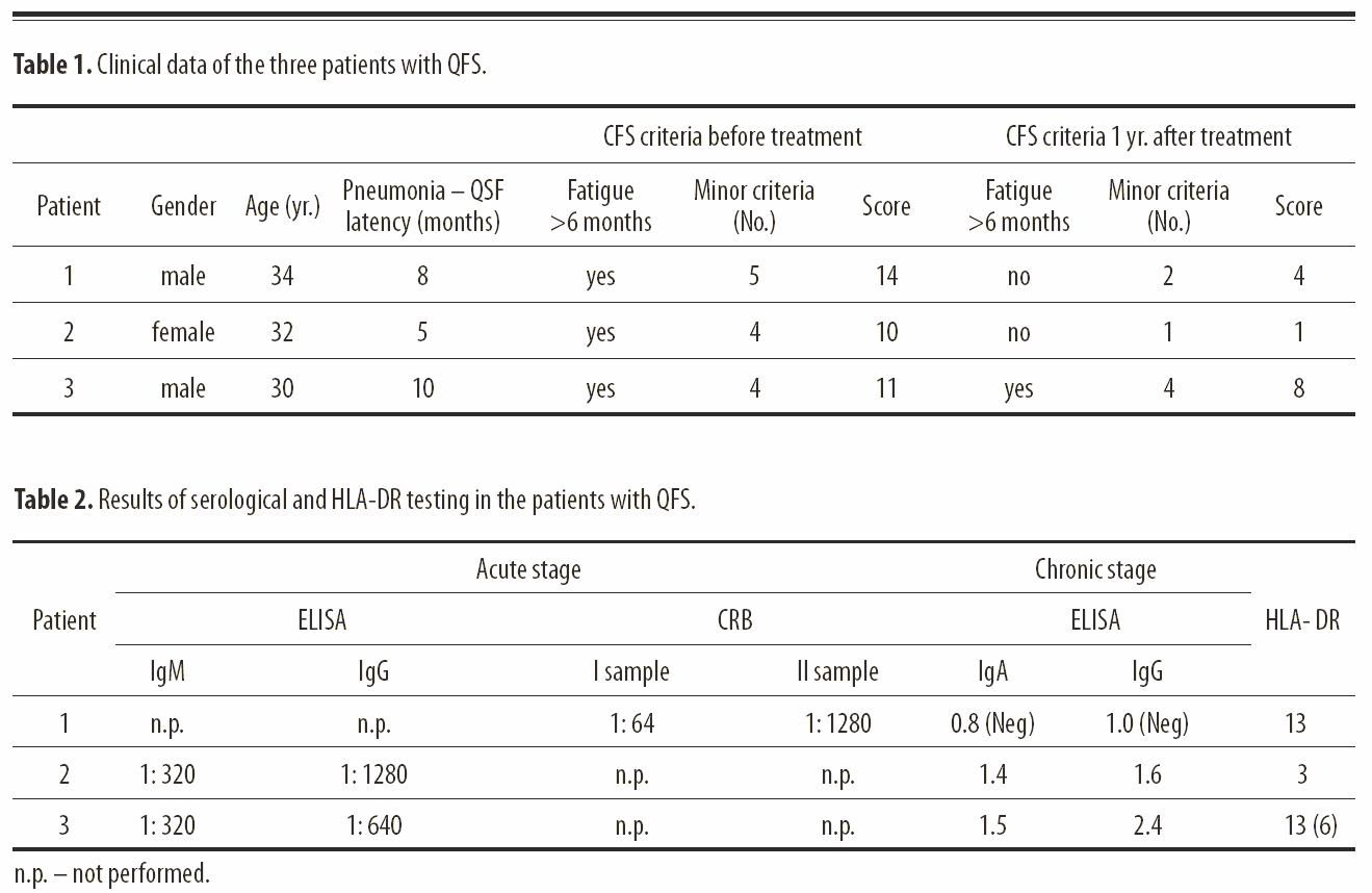
considered favorable (Table 1).
CASE REPORTS
Case 1
A 34-year-old male shopkeeper
with atypical pneumonia caused by
Coxiella burnetii was treated
at the Department for Pulmonary Diseases in February 2000. He did
not have any serious illness before he caught Q fever. He arrived
from a rural area where Q fever is endemic. Laboratory results
showed an erythrocyte sedimentation rate (ESR) of 72 mm/hour, while
the other hematological and biochemical parameters showed no
abnormalities. The patient received a combination of erythromycin
4×500 mg/day p.o. and gentamycin 1×240 mg/day i.v for two weeks. The
clinical response was good. A control chest x-ray was normal. The
etiology was confirmed by the complement-binding reaction (CBR),
which showed a titer for Coxiella burnetii of 1:64. A repeat
CBR for Coxiella burnetii after six weeks was 1:1024.
During follow-up
within the year 2000, the patient complained of disrupted sleep,
morning fatigue, intense headache, prolonged fatigue lasting more
than 24 hours after physical work, muscle pain, and persistent
low-grade fever. Transthoracic heart ultrasound was normal. Serology
for the phase I and phase II replication cycle of Coxiella
burnetii did not confirm chronic infection (Table 2). After a
one-year duration of symptoms, nine months of treatment with
ciprofloxacin (2×500 mg/day p.o.) and doxycycline (2×100 mg/day
p.o.) was instituted. The muscle pain and low-grade fever
disappeared after this therapy, but the mild headache persisted.
Therefore, in January 2002 a lumbar tap was performed. Cytology and
biochemistry of CSF showed no abnormalities. The CSF sample was
tested for Coxiella burnetii using an indirect immunofluorescence assay and the result was negative. The patient still has
low intensity headache and he suffers from fatigue after physical
activity, but it disappears after half an hour of rest. He has
returned to work, but has changed his job from shopkeeper to
watchman. He now suffers from hyperlipidemia and does not show
criteria for chronic fatigue syndrome (Table 1).
Case 2
A 30-year-old male professional soldier with
interstitial pneumonia was treated at the Department for Pulmonary
Diseases of the Clinical Hospital of Split in February 2004.
In the acute phase of illness his ESR was 46
mm/hour, while other hematological test results were normal. Blood
chemistry values were normal with the exception of AST 62 U/l
(normal range: 0–29) and ALT 54 U/l (normal range: 0–30). After two
weeks of treatment with doxycycline, pulmonary infiltrates resolved
and hematological and other laboratory results were all within the
normal ranges. IFA for Coxiella burnetii revealed positive
IgM 1:64 and IgG 1:320 in a first and IgM 1:320 IgG 1:640 one month
later in a second serum sample. Four months later the patient
started complaining of fatigue, disrupted sleep, headaches, and
muscle and joint pain. Therapy with corticosteroids was introduced
and continued for one month without success. In January 2005 the
patient was admitted to the Department for Infectious Diseases, and
his routine hematological and biochemical tests were within
physiological limits. ELISA for Epstein-Barr virus, cytomegalovirus,
HIV, and Toxoplasma gondii were doxycycline for two weeks
with a good clinical response, and her chest x-ray after two weeks
confirmed complete regression of pulmonary infiltrations. An
indirect immunofluorescence test (IFT) in the acute stage of the
disease showed positive IgM (titer: 1:160) and IgG (titer: 1:640)
for Coxiella burnetii. Repeated serology one month later
showed IgM 1:320 and IgG 1:1280. After she had felt well for two
months, she started experiencing pain in her neck. Six months later,
in August 2003, in addition to the neck pain she began to suffer
from insomnia, headache, sweating, and fatigue, which did not
resolve after sleep. The symptoms persisted for 12 months.
She was admitted to the Department for Infectious
Diseases again in October 2004. Repeated hematological and
biochemical results were within physiological values.
Electromyography of the upper and lower extremities showed no
abnormalities and transthoracic and transesophageal heart ultrasound
showed no signs of endocarditis.
Rheumatoid factor, antinuclear antibodies, and antimitochondrial
antibodies as well as serology for Epstein-Barr virus,
cytomegalovirus, and toxoplasmosis were negative. Anti-HIV and
hepatitis B and C markers were also negative, and thyroid hormones
were within normal ranges. Paired serum samples in ELISA for
Coxiella burnetii showed positive phase I IgA and IgG antibodies
(Table 2). The therapy included ciprofloxacin (2×500 mg/day p.o.)
for two months followed by doxycycline (2×100 mg/day p.o.) for four
months. The result of the six months of treatment was regression of
symptoms, with only a minor headache persisting. She is now capable
of doing all her housework and does not fulfill the criteria for CFS
(Table 1).
Case 3
A 30-year-old male professional soldier with
interstitial pneumonia was treated at the Department for Pulmonary
Diseases of the Clinical Hospital of Split in February 2004.
In the acute phase of illness his ESR was 46
mm/hour, while other hematological test results were normal. Blood
chemistry values were normal with the exception of AST 62 U/l
(normal range: 0–29) and ALT 54 U/l (normal range: 0–30). After two
weeks of treatment with doxycycline, pulmonary infiltrates resolved
and hematological and other laboratory results were all within the
normal ranges. IFA for Coxiella burnetii revealed positive
IgM 1:64 and IgG 1:320 in a first and IgM 1:320 IgG 1:640 one month
later in a second serum sample. Four months later the patient
started complaining of fatigue, disrupted sleep, headaches, and
muscle and joint pain. Therapy with corticosteroids was introduced
and continued for one month without success. In January 2005 the
patient was admitted to the Department for Infectious Diseases, and
his routine hematological and biochemical tests were within
physiological limits. ELISA for Epstein-Barr virus, cytomegalovirus,
HIV, and Toxoplasma gondii were negative. Transthoracic and transesophageal heart
ultrasound showed no signs of endocarditis. Ultrasound of abdomen
was also normal. Rheumatoid factor, antinuclear antibodies, and
antimitochondrial antibodies were negative. Biphasic ELISA test for
Coxiella burnetii showed positive IgA antibodies in phase I (Table
2). After completing three months of antibiotic treatment with
doxycycline, the patient still had fatigue, disrupted sleep,
headaches, and muscle and joint pain. He still fulfills the criteria
for CSF, cannot go back to work, and awaits realization of his
retirement (Table 1).
DISCUSSION
Three patients with diagnoses of chronic fatigue
syndrome after Q fever are described. Positive IgA antibodies for
phase I of the Coxiella burnetii growth cycle suggest the
possibility of chronic infection and the presence of Coxiella
burnetii in macrophages [7]. Two of the patients described in
this study had positive IgA antibodies for phase I of the
Coxiella burnetii growth cycle and serology which was consistent
with chronic Coxiella burnetii infection, while patient No. 1
had negative serology for chronic Coxiella burnetii infection
(Table 2).
As there are no clinical signs
or laboratory tests that could be taken as definite proof of CFS,
the disease is diagnosed based on the patients’ symptoms and by
excluding other diseases with similar symptoms [8]. In the last ten
years, Q fever has been included in a group of diseases that are
associated with the development of CFS after the acute phase of
illness [7].
A recent article by Hickie et al. suggests
that post-infective fatigue syndrome can occur after clinical
infection by several different viral and non-viral microorganisms.
The authors suggest that the CFS phenotype was stereotyped and
occurred with similar incidence after Epstein-Barr virus, Q fever,
and Ross River virus infection. The occurrence of CFS was predicted
in the highest degree by the severity of the acute infection [12].
All our patients had moderately severe acute illness. Helbig and
associates suggest a genetic predisposition for CFS[13]. Analyzing
patients who had Q fever in England, Ayres and associates
established that long persistence of fatigue, increased sweating,
blurred vision, and shortening of breath are manifested more
commonly in the group of patients that suffered from Q fever than in
the control group [14]. Similar results were obtained by Marmion’s
et al. [15] while comparing slaughterhouse workers who had Q fever
with a serologically negative control group. Fatigue, headache,
disrupted sleep, and muscle and joint pain were significantly more
frequent in the group of workers with previous Q fever. Ayres [14]
associated shortness of breath in patients after Q fever with
possible myocardial lesions after
Coxiella burnetii
infection, that were first
referred to by Maisch in 1986 [16]. Lovey et al. [17] established a
higher incidence of cardiovascular diseases in patients who had Q
fever in comparison with a control group. Later studies by Ayres et
al. did not show any significant difference in cardiological
measurements that would suggest cardiomyopathy or other heart
diseases when comparing a group with CFS after acute Q fever and a
group without symptoms of CFS [18].
Thomas et al. did not find any significant
differences in the frequencies of fatigue, depression, and lack of
concentration between individuals with positive antibodies for
Coxiella burnetii and serologically negative individuals. The
imperfection of this study was that it included all Q-feverseropositive
individuals without differentiation between patients who had
asymptomatic and those who had symptomatic acute Q fever, as well as
the fact that the study was done on a relatively healthy population
with little neuropsychiatric morbidity [19]. Although Marmion et al.
suggested that the diagnosis of QFS does not require serological
criteria for chronic Q fever, low serological titers against C.
burnetti were
associated with chronic fatigue syndrome by Penttila et al. [15,11].
It is therefore not clear if patients with symptoms of CFS and
positive serology of chronic Q fever, but lacking other clinical
manifestations of chronic Q-fever such as endocarditis or osteitis,
as described in the cases 2 and 3 of this paper, should be included
in this syndrome. We therefore believe that patients with CFS
criteria, positive phase I serology, and without other clinical
manifestations of chronic Q fever should be diagnosed as QFS.
Finally, is there any
usefulness of antibiotic therapy of post-Q-fever CFS? The results of
antibiotic therapy in patients presented in this paper were
conflicting: in two cases the symptoms diminished, while the third
patient continued to complain of CFS symptoms. These results are
based on their clinical findings, before and after the therapy, as
well as a questionnaire investigation. Up to now, there are two
studies investigating the outcome of QFS therapy. Arashima et al.
conducted treatment with minocycline for a period of three months in
twenty patients with QFS. The result was satisfactory, and in all
patients fatigue resolved, while seven patients with positive PCR
test for
Coxiella burnetii turned negative [20]. The limitation of
this study is the absence of a placebo control group. One year
later, Iwakami et al. Studied the effects of three months of
antibiotic therapy in patients with post-Q-fever CFS. Although they
became negative for C. burnetii
DNA, in contrast to Arashima’s study no
improvement of their symptoms was observed [21]. Another anecdotal
attempt was the treatment of three-year-old girl with post-Q-fever
CFS with interferon-g after unsuccessful antibiotic therapy [22].
The idea for such therapy was
based on the knowledge that interferon-g induces the killing of
monocytes infected with
Coxiella burnetii. The result
of treatment was satisfying and encouraging for further
investigations. Although Vissar et al. [23] accentuated the
diversity of the immune response of peripheral mononuclear cells in
patients with CFS after stimulation with dexamethasone, our patient
treated with corticosteroids did not experience amelioration of his
symptoms.
CONCLUSIONS
Our case series of patients from southern Croatia, where Q fever
is endemic, is in concordance with more detailed data presented in
the past from other areas of the world. Therefore we can conclude
that a substantial number of patients develops CFS after acute Q
fever in spite of appropriate antibiotic therapy during acute
infection. The results of prolonged antibiotic therapy in such the
patients are inconsistent. Efforts to establish diagnostic criteria
as well as therapeutic recommendations for post-Q-fever CFS require
further investigation.
REFERENCES:
|
1. |
Brouqui P, Marrie TJ, Raoult D: Coxiella In: Murray PR, Baron
EJ, Jorgenson JH, Phaler MA, Yolken RH (eds.) Manual of clinical
microbiology. 8th ed. Washington D.C, ASM press; 2003; 1030–36 |
|
2. |
Marrie TJ, Raoult D:
Coxiella burnetii (Q fever). In: Mandell GL, Douglas JE, Bennett JE
(eds.) Principles and Practice of Infectious Diseases. 6th ed. New
York: Churchill Livingstone, 2005; 2296–302 Med Sci Monit, 2007;
13(7): CS88-92 Ledina D et al – Chronic Fatigue Syndrome and Q fever
CS91 |
|
3. |
Punda-Polić
V, Radulović S: Sero-survey of Q fever
in the north-western part of Bosnia and Herzegovina. Croat Med J,
1997; 38: 345–47 |
|
4. |
Medić
A, Dželalija B, Punda Polić V et al: Q fever epidemic
among employees in a factory in the suburb of Zadar, Croatia. Croat
Med J, 2005; 46(2): 315–19 |
|
5. |
Lukšić
B, Punda-Polić
V, Ivić I et al: Clinical and
epidemiological features of hospitalized acute Q fever cases from
Split-Dalmatia County (Croatia), 1985–2002. Med Sci Monit, 2006;
12(3): CR126–31 |
|
6. |
Puljiz I, Kuzman I, Daković-Rode
O: Clinical and epidemiological characteristics of Q fever in
hospitalised patients. Infektol. Glasn, 2005; 25: 75–80 |
|
7. |
Raoult D, Marrie TJ, Mege JL:
Natural history and pathophysiology of Q fever. Lancet Infect Dis,
2005; 5: 219–26 |
|
8.
|
Fukuda K, Straus SE, Hickie
I et al: The Chronic fatigue syndrome: A Comprehensive Approach to
its Defi nition and Study. Ann Intern Med, 1994; 121: 953–59
|
|
9. |
Engelberg NC: Chronic
Fatique Syndrome. In: Mandell GL, Douglas JE, Bennett JE (eds.)
Principles and Practice of Infectious Diseases. 6th ed. New York:
Churchill Livingstone; 2005; 120–25 |
|
10. |
Korzon M, Bukowska W,
Szlogatys-Sidorkiewicz A: Chronic fatigue syndrome. Med Sci Monit,
1998; 4(2): 388–92 |
|
11. |
Penttila IA, Harris RJ,
Storm P et al: Cytokine dysregulation in the post-Q-fever fatigue
syndrome. Q J Med, 1998; 91: 549–60 |
|
12. |
Hickie I, Davenport T,
Wakefield D et al: Post-infective and chronic fatigue syndromes
precipitated by viral and non-viral pathogens: prospective cohort
study. BMJ, 2006; 333(7568): 575–81 |
| 13. |
Helbig KJ, Heatley SL,
Harris RJ et al: Variation in immune response genes and
chronic Q fever. Concepts: Preliminary test with post-Q
fever fatigue syndrome. Genes Immun, 2003; 4: 82–85
|
|
14. |
Ayres JG, Flint N, Smith EG et al: Post-infection fatigue syndrome following Q fever. Q J
Med, 1998; 91: 105–23 |
|
15. |
Marmion BP, Shannon M,
Maddocks I et al: Protracted debility and fatigue after acute Q
fever. Lancet, 1996; 347: 977–78 |
|
16. |
Maisch B: Rickettsial
perimyocarditis-a follow up study. Heart Vessels, 1986; 2: 55–59 |
|
17. |
Lovey P-Y, Morabia A,
Bleed D et al: Long term vascular complication of Coxiella burnetii
infection in Switzerland: cohort study. Br Med J, 1999; 319: 284–86 |
| |
18. Ayres JG, Wildman M,
Groves J et al: Long-term Follow-up of patients from the 1989 Q
fever outbreak: no evidence of excess cardiac disease in those with
fatigue. Q J Med, 2002; 95: 539–46 |
|
19. |
Thomas HV, Thomas DR,
Salmon RL et al: Toxoplasma and Coxiella infection and psychiatric
morbidity: A retrospective cohort analysis. BMC Psychiatry, 2004; 4:
326–29 |
|
20. |
Arashima Y, Kato K,
Komiya T et al: Improvement of Chronic Nonspecific symptoms by
long-term minocycline treatment in Japanese patients with
Coxiella burnetii infection considered to have post-Q fever
fatigue syndrome. Intern Med, 2004; 43: 49–54
|
|
21. |
Iwakami E, Arashima Y,
Kato K et al: Treatment of chronic fatigue syndrome with
antibiotics: Pilot study assessing the involvement of Coxiella
burnetii infection. Intern Med, 2005; 44: 1258–63 |
|
22. |
Yutaka M, Hiroshi W, Tomoki T et al:
Intractable Q fever treated with recombinant gamma interferon. Pediatr Infect Dis, 2001; 20: 547–57 23. Visser J, Blauw B, Hinlopen
B et al: CD4 T lymphocytes from patients with chronic fatigue
syndrome have decreased interferon-g production and increased
sensitivity to dexamethasone. J Infect Dis, 1998; 177:451–54 |

Last Updated:
23/01/2011
|